
Structural Heart (TAVI & TEER) | Dr. Naveen Bhamri
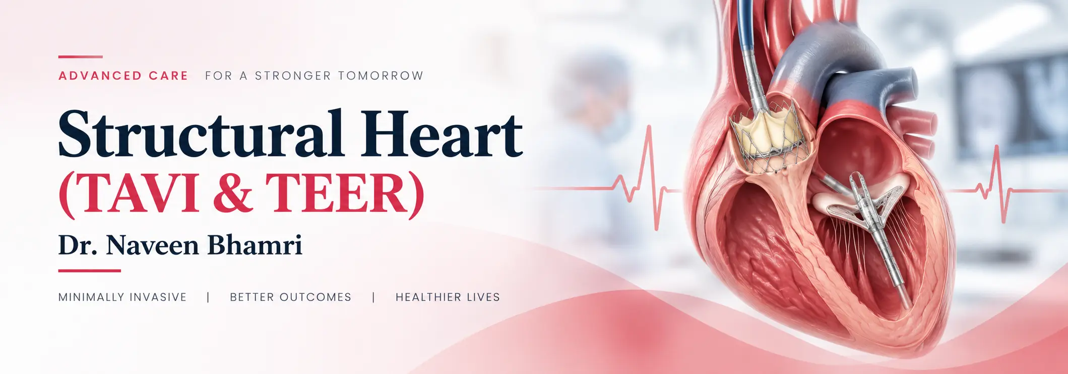
Structural heart disease affects the heart’s valves or other structures and can interfere with how efficiently the heart pumps blood. TAVI (Transcatheter Aortic Valve Implantation) and TEER (Transcatheter Edge-to-Edge Repair) are minimally invasive, catheter-based treatments used for selected patients with significant valve disease. They may be considered when symptoms are affecting daily life or when conventional surgery may not be the most suitable option. Dr. Naveen Bhamri is regarded as the best Structural heart doctor in Delhi and the best cardiologist at Max-BLK Hospital in Shalimar Bagh, with specialized expertise in heart valve treatment, including TAVI, as part of his interventional cardiology practice.
What Are TAVI and TEER?
When Are TAVI or TEER Recommended?
The decision depends on the valve involved, severity of disease, symptoms, heart function, overall health, and surgical risk.
TAVI may be considered when a patient has:
- Severe aortic valve stenosis
- Breathlessness, chest discomfort, dizziness, or reduced exercise tolerance
- Symptoms affecting routine activities
- An increased risk or concern associated with conventional valve surgery
- A valve condition where catheter-based replacement is considered appropriate
TEER may be considered in selected patients with:
- Significant mitral or tricuspid valve regurgitation
- Breathlessness or reduced exercise capacity related to valve leakage
- Persistent symptoms despite appropriate medical treatment
- Increased surgical risk or circumstances where a catheter-based approach may be preferable
Not every patient with valve disease requires an intervention. Treatment should follow detailed clinical and imaging assessment.
Symptoms or Signs You Should Not Ignore
Valve disease can develop gradually, and symptoms may initially be mistaken for aging or reduced fitness. Watch for:
- Breathlessness during activity or at rest
- Unusual tiredness or reduced stamina
- Chest discomfort or pressure
- Dizziness or fainting
- Palpitations
- Swelling of the legs or ankles
- Difficulty performing activities that were previously comfortable
New, worsening, or severe symptoms should be medically evaluated rather than self-managed.
How Are Structural Heart Conditions Diagnosed?
Evaluation may include:
- Clinical examination and medical history
- Electrocardiogram (ECG)
- Echocardiography
- Transesophageal echocardiography (TEE), when required
- CT scan for procedural planning
- Cardiac catheterization
- Assessment of heart function and valve severity
- Evaluation of surgical and procedural risk
For TAVI and TEER, imaging plays an important role in determining whether the anatomy is suitable for a catheter-based procedure.
TAVI (Transcatheter Aortic Valve Implantation)
TAVI is a minimally invasive procedure used to replace a diseased aortic valve without conventional open-heart valve surgery. The procedure generally involves:
Pre-procedure assessment: Detailed echocardiography and CT imaging help assess the aortic valve, heart anatomy, and suitable access route.
Catheter access: A thin catheter is introduced through a blood vessel, commonly through the femoral artery.
Valve positioning: The replacement valve is guided through the catheter and positioned inside the diseased aortic valve.
Valve deployment: The new valve is expanded and secured in place, taking over the function of the narrowed native valve.
Final assessment: Imaging and pressure measurements are used to assess valve function and check for complications.
Recovery: Patients are monitored after the procedure, with recovery generally focused on early mobilization and follow-up.
TEER (Transcatheter Edge-to-Edge Repair)
TEER is a catheter-based procedure used in selected patients with significant valve regurgitation. It works by bringing portions of the valve leaflets together to reduce backward blood flow.
Imaging assessment: Detailed echocardiography, often including transesophageal echocardiography, is used to assess valve anatomy and determine suitability.
Catheter access: A catheter is introduced through a blood vessel and guided towards the heart.
Device positioning: A specialized repair device is guided to the affected valve under continuous imaging guidance.
Leaflet approximation: The device brings selected valve leaflets together, helping reduce the amount of blood leaking backward.
Assessment: The valve is reassessed to determine the reduction in regurgitation and ensure satisfactory function.
Post-procedure monitoring: The patient is monitored following the procedure, with follow-up planned based on individual clinical needs.