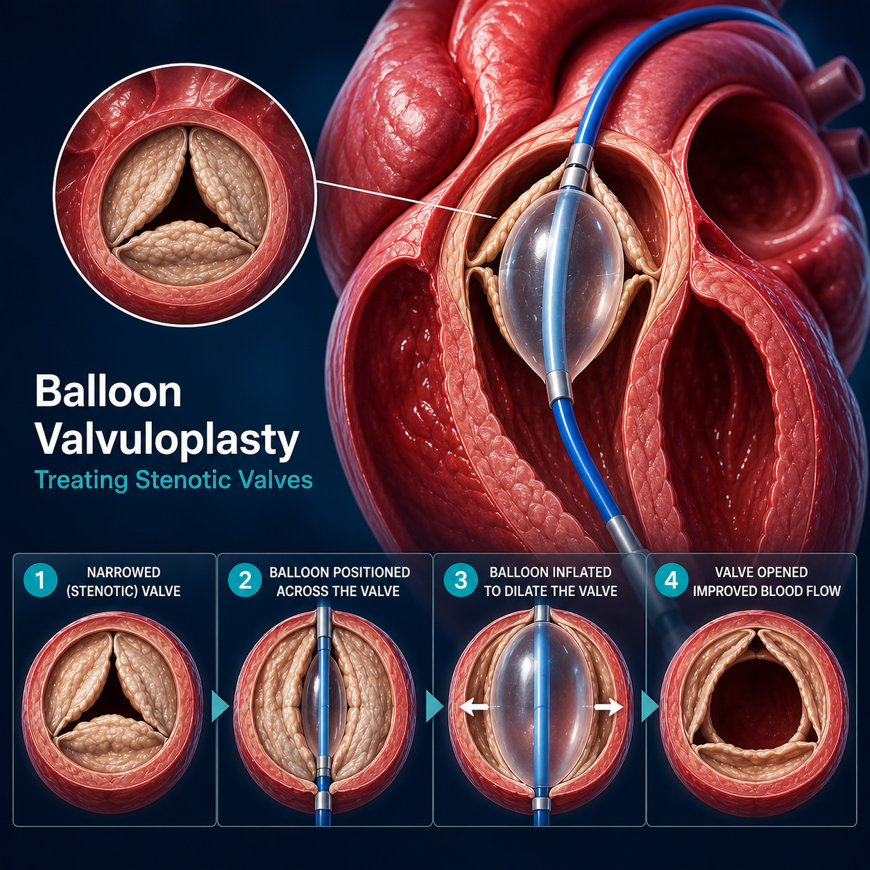
Balloon valvuloplasty is a percutaneous (through the skin) procedure, meaning it does not require opening the chest. It is often used for the mitral, aortic, or pulmonary valves.
1. Access and Navigation
A thin, flexible tube (catheter) is inserted through a blood vessel, usually in the groin. Using real-time X-ray imaging (fluoroscopy), the cardiologist guides the catheter up into the heart and positions it precisely within the narrowed valve.
2. The Dilation Process
Placement: A specialized deflated balloon at the tip of the catheter is placed across the stiff valve leaflets.
Inflation: The doctor quickly inflates the balloon. The force of the inflation stretches the valve opening and separates the leaflets that have become fused or stuck together.
Verification: The balloon is deflated and inflated several times to ensure the opening is sufficiently widened.
3. Completion
Once the valve is sufficiently opened, the balloon is deflated and removed along with the catheter. The valve can now open more freely, restoring better blood flow and reducing the workload on the heart muscle.